 Ion-exchange chromatography (IEX) is a process that allows the separation of molecules based on their charge. It can be used for almost any kind of charged molecule including large proteins such as haemoglobin.
For the detection of haemoglobinopathies, haemoglobin samples are first introduced via an autosampler into a sample loop of known volume. A buffered aqueous solution known as the mobile phase then carries the sample from the loop onto a column. This column contains an ion-exchange resin bound to a silica gel support has been equilibrated with respect to pH and ionic strength.
Separation of hemoglobin species is accomplished through the use of a gradient between two mobile phases with differences in salt concentration and pH, with high pressure pumps transfering mobile phases through the analytical column. Such physical characteristics as surface charge, as well as the presence of hydrophilic and hydrophobic groups determine the rate each haemoglobin species migrates through the column.
Upon elution from the column, sample components pass through the spectrophotometric detector where detection occurs at a wavelength of 413nm ± 2nm. Following elution of all hemoglobin species, original conditions are re-established prior to the injection of the next sample. All critical events including sample injection, reagent flow rates, and composition are carefully timed to provide maximum reproducibility.
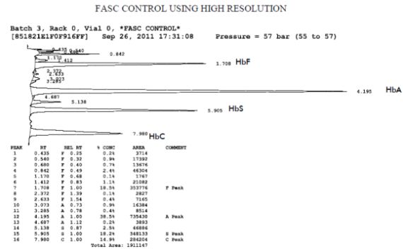
All processes are electronically controlled through the computer. The computer processes the signal from the detector and calculates the retention time and percent concentration of each peak. Integration is by peak area in millivolt-seconds.
As the sample is chromatographed, it is displayed on the monitor in real time. The computer produces printed reports with the sample identification information, date and time, followed by the chromatogram with retention times indicated at the apex of each peak. An example peak summary report is illustrated below.
